

BARRETT’S ESOPHAGUS FISH
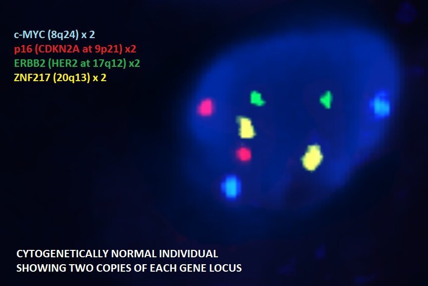
Negative Barrett’s Esophagus

Positive Barrett’s Esophagus
OmniPathology is proud to offer FISH testing for Barrett’s esophagus (BE) performed on esophageal biopsies. Testing on biopsies has many advantages over esophageal brushes.
BE FISH identifies cytogenetic abnormalities that are associated with cellular progression toward adenocarcinoma. Studies showed that the combination of histology and four-probes FISH has superior sensitivity detecting low grade dysplasia LGD, high-grade dysplasia (HGD) and adenocarcinoma than other techniques including cytology and digital image analysis.
INTENDED USE:
This four-probes FISH assay is performed on biopsies showing indefinite for dysplasia (ID), low-grade dysplasia (LGD) and high-grade dysplasia (HGD) to augment the morphologic assessment.
OMNI BE FISH HELPS THE PATHOLOGIST ACHIEVE THE FOLLOWING:
-
Current guidelines require confirmation of HGD by sending the case for a second opinion by an expert pathologist. Using FISH obviates the need for a second review.
-
Retrospective analysis of patients with HGD showed that there is increased risk of progression to adenocarcinoma when polysomy is present in four of 100 cells.
-
FISH analysis, when performed on cases of ID and LGD, can predict the presence of HGD somewhere else.
ADVANTAGES OF FISH TESTING ON BIOPSIES VS CYTOLOGY BRUSHES:
One of the most critical components of the BE diagnosis are anatomical landmarks. In the United States, the diagnosis of BE requires the identification of intestinal metaplasia (goblet cells) within the anatomic esophagus, which is above the lower esophageal sphincter (LES). Intestinal metaplasia at the gastroesophageal junction (GEJ) or below the LES is not diagnostic of the BE. That makes biopsies superior to brushes because brush specimens are obtained by going up and down across the GEJ, recovering cells from GEJ and below the LES. Furthermore, dysplasia assessment in cytology specimens is less accurate than biopsies due to lack of the architecture in cytology specimens. In biopsies, changes that do not reach the surface can be classified as indefinite for dysplasia (ID). Such architectural context is not available in cytology samples, which results in difficulty in grading dysplasia. In fact, the diagnostic category of indefinite for dysplasia is not a cytological entity.
REFERENCE:
Poneros JM, Faye AS, Barr Fritcher EG, et al. A Multicenter Study of a Fluorescence In Situ Hybridization Probe Set for Diagnosing High-Grade Dysplasia and Adenocarcinoma in Barrett's Esophagus. Dig Dis Sci. 2017;62(5):1216–1222. doi:10.1007/s10620-017-4517-y