
OMNI Expands Digital Pathology Capabilities to Advance Gastrointestinal Diagnostics
At OmniPathology, innovation is not simply about adopting new technology. It is about finding practical ways to use technology to improve diagnostic quality, efficiency, and ultimately patient care.
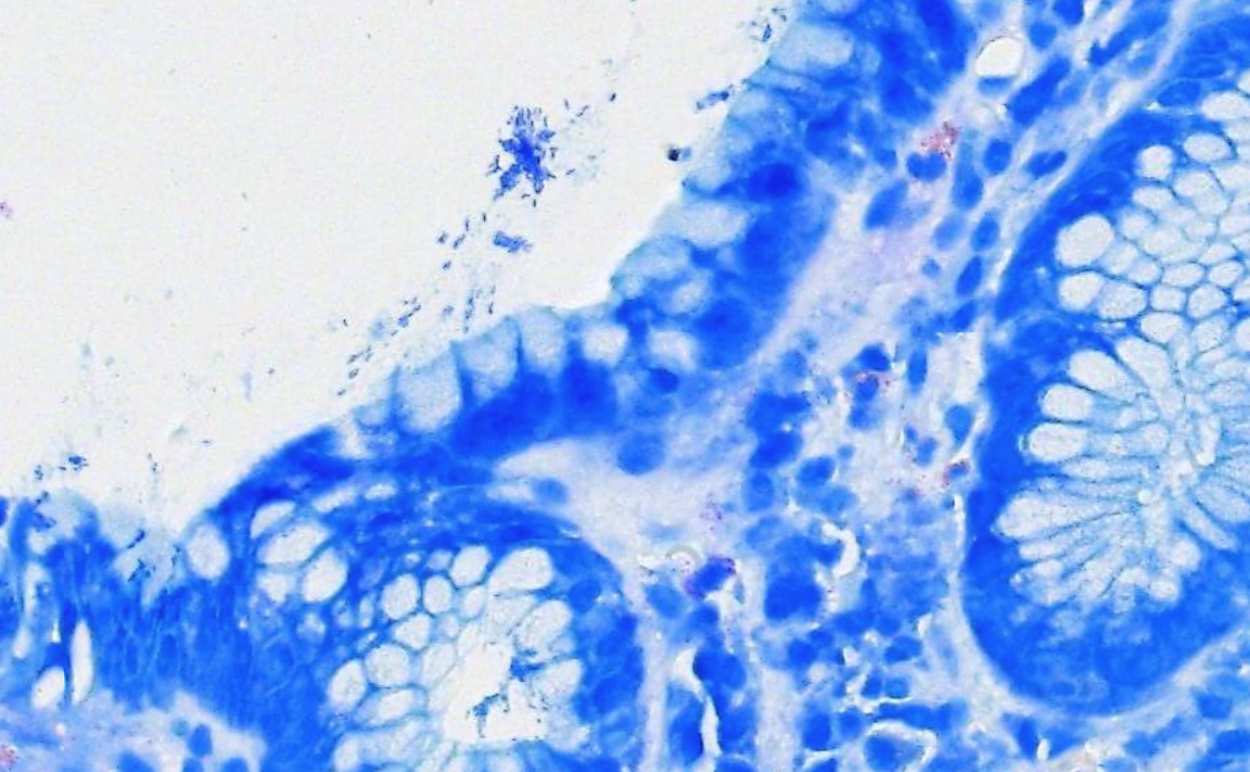
As part of our continuing expansion of digital pathology, OmniPathology continues to pursue solutions to the most common challenges. One of the industry’s challenges is the digital detection of micro-organisms, particularly in gastrointestinal (GI) pathology. More specifically, the digital detection of Helicobacter pylori (H. pylori), Giardia duodenalis, and spirochetes. Because of their small size compared to gastric and intestinal cells, they are often difficult to clearly resolve with high-throughput digital scanners.
A Small Organism with Significant Clinical Importance
OmniPathology evaluates a high volume of gastric biopsies every day. One of the important diagnostic questions in these specimens is whether H. pylori is present.
This small bacterium is associated with chronic active gastritis and peptic ulcer disease and is a major risk factor for gastric cancer. Identifying H. pylori accurately and efficiently is therefore an important component of the evaluation of many gastric biopsy specimens. The use of histochemical staining techniques, such as Giemsa, has been an established and efficient method to clearly identify H. pylori for years. Unfortunately, resolving H. pylori using high-throughput digital scanners has been challenging, and many labs have resorted to modifying their workflows and adopting expensive immunohistochemistry stains to identify the pathogens.Solving a Digital Pathology Challenge
As pathology laboratories transition from glass slides to digital images, it is important that solutions to challenges not result in workflow disruption or increased cost. For a high-volume GI pathology laboratory, even seemingly small workflow changes can have a meaningful operational and financial impact.
For OmniPathology, this represents an important criteria toward creating a more comprehensive digital workflow specifically designed around the realities of GI pathology.
Building a More Complete Digital GI Pathology Workflow
As a leading organization in GI pathology, OmniPathology's goal is to have full digitization of its GI pathology slides including challenging specimens while supporting the high-throughput requirements of our laboratory.
By incorporating specialized imaging capabilities into our existing digital pathology environment, Omni continues to move toward a future in which digital pathology is not simply an alternative way to view a glass slide, it becomes an integral part of how pathology is performed.
Our goal is to combine advanced imaging, pathology expertise, laboratory science, and innovative technology to create digital workflows that are practical, scalable, and focused on delivering the highest quality diagnostic information to physicians and their patients.